English
+37494840986 / +37499016063

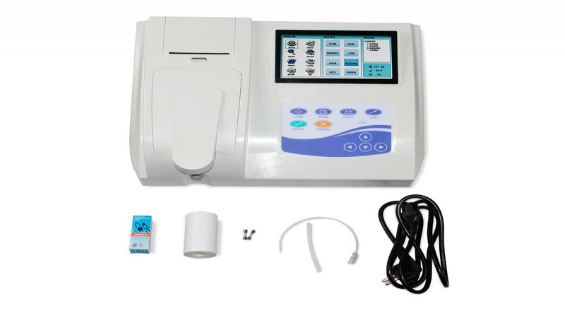
Laboratory equipment
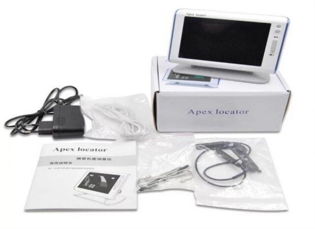
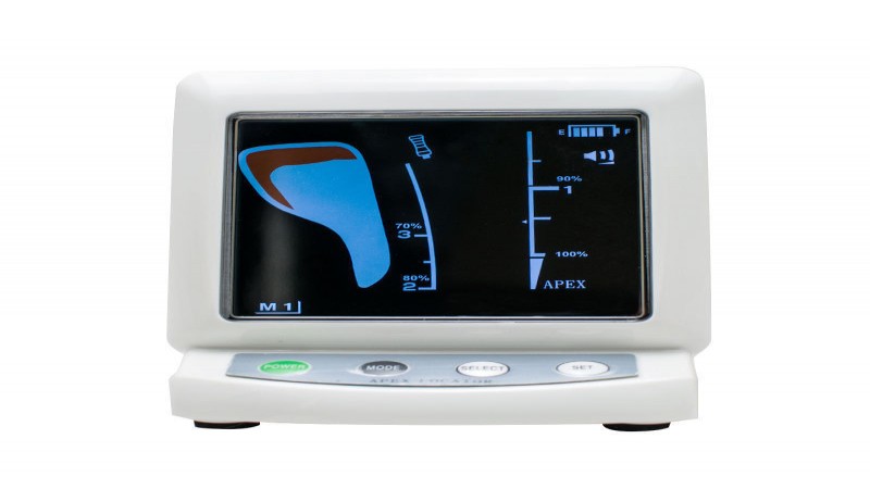
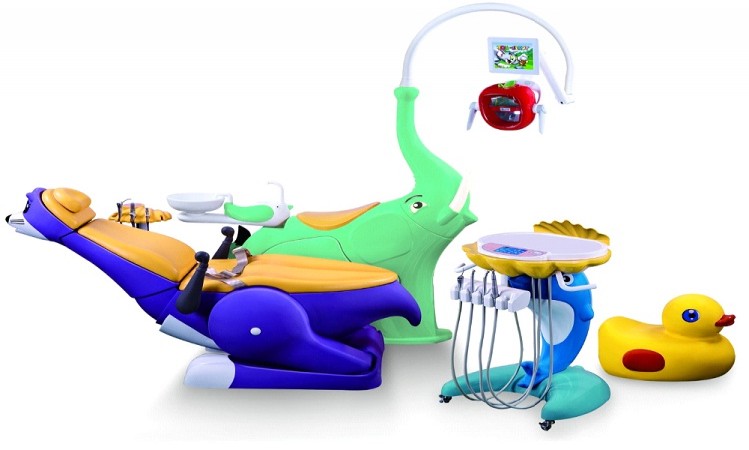
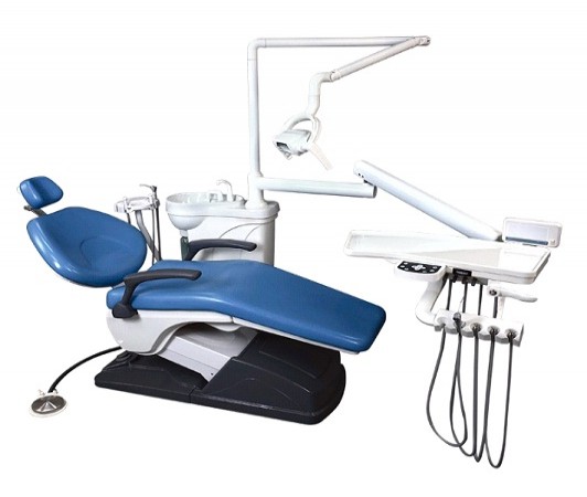
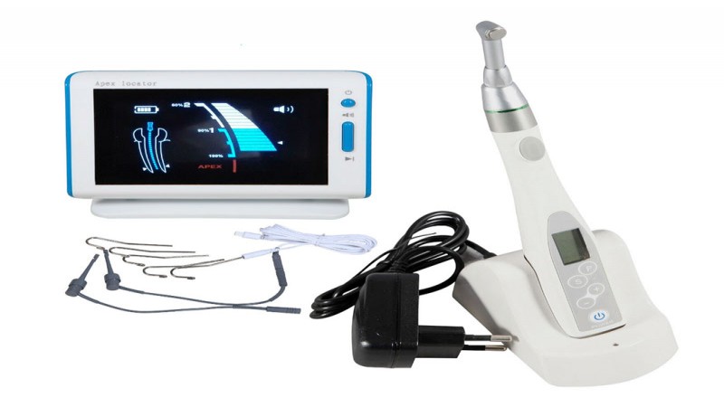
Dental equipment
ENT equipment
Ophthalmological equipment
Sterilization and disinfection
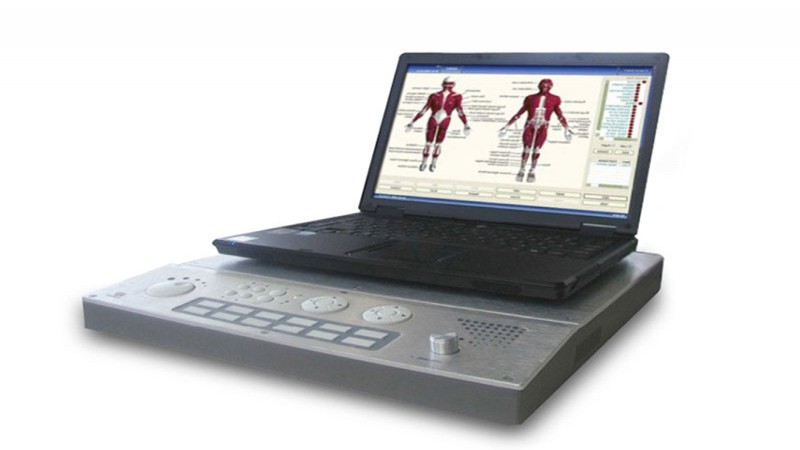
Diagnostic equipment
Gynecological equipment
Dermatological equipment
Cosmetology equipment
Surgical Equipment
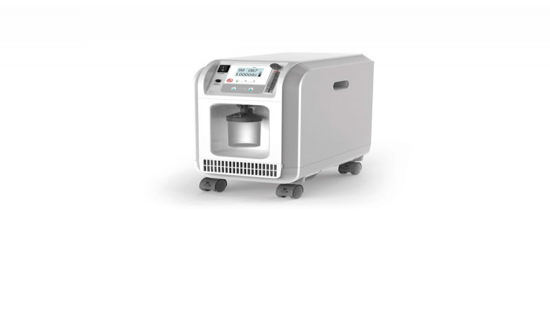
Pulmonary equipment
Computed tomography CT